Friday, December 10, 2010

PDA ligation controversies


Well, that was another excellent joint neonatal-Pediatric surgery conference at WCHOB. The subject was PDA and the discussion included basic PDA physiology, morbidity, treatment strategies, post ligation syndrome, and most importantly looked at the evidence (or lack of) supporting the need for PDA ligation.

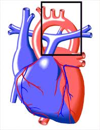
The discussion began with the general principles of PDA physiology, including the normal role of prostaglandins produced by the placenta and the high intraluminal blood pressure in the ductus (secondary to high pulmonary vascular resistance) in keeping the ductus patent in utero. Normal term delivery reverses these factors and results in closure of the ductus initially by smooth muscle constriction then then anatomic remodeling.

The clinical consequences of a PDA are a result of the left to right shunt, and subsequent change in blood flow to vital organs with subsequent metabolic acidosis, increased risk of IVH and NEC secondary to diastolic steal, and pulmonary vascular disease due to continuous dilation of pulmonary vessels during diastole.

The main strategy for prophylaxis and treatment is the administration of Indomethacin, and when that fails, surgical ligation.

The first issue that comes up is the evidence for the need for the prophylaxis and treatment of PDA.

The literature supports the benefit of the prophylactic use of indomethacin for it's effectiveness of PDA closure, decrease the need for surgical ligation, decrease in the incidence and severity of pulmonary hemorrhage, and decrease in incidence of grade 3 and 4 IVH.

Indomethacin, however, is not innocuous. It can cause a transient change in renal function, increases the risk of GI perforation when administered simultaneously with steroids. It was not found to have any neurodevelopemental effects.

So why should a PDA be treated? A study looking at infants <1500 g showed an 8 fold increase in mortality in the presence of a PDA.

When it came to the subject of surgical ligation of PDA, the data was much less convincing and somewhat troubling. Studies not only shed doubt on the proposed rapid improvement of cardiovascular parameters (Raval et al. J Ped Surg 2007.42(1):69), some actually showed that surgical ligation may be associated with an increased risk of BPD, severe ROP, and neurosensory impairment in ELBW infants (Kabra et al. J Peds 2007;150:229).

Finally, the subject of post ligation cardiac dysfunction was addressed. This results from ligation of the PDA, which results in a sudden switch from volume to pressure overload on the heart which was, as a baseline, subjected to impaired coronary perfusion (form diastolic steal with a PDA), and preexisting pulmonary edema and cardiac failure. Post ligation cardiac dysfunction presents with severe hypotension, failure of oxygenation, and myocardial dysfunction. The hemodynamic profile shifts from pre ligation state of a high preload/low after load and myocardial ischemia from decreased diastolic perfusion, to post ligation state of a sudden increase in after load, decreased LV end diastolic volume, and decreased LV output.

Importantly, the extent of post ligation dysfunction depends on pre ligation hemodynamic, with some authors supporting the prophylactic use of milrinone when the pre ligation LV output is < 200 ml/kg/minute.

As to the management of post ligation cardiac dysfunction, the strategy is to use inotropes that do not increase after load (Milrinone/dobutamine), optimize oxygen carrying capacity, and fluid management.

The upshot on PDA interventions was that prophylactic ligation does not improve outcome, indomethacin prophylaxis decreases IVH and need for PDA ligation, indomethacin prophylaxis does not improve neurodevelopemental outcome in survivors, earlier Indomethacin treatment is associated with higher rates of DA closure. As to the ideal timing of medical or operative closure, that remains to be clarified.

With the current available data, many questions remain unanswered:

can we identify infants whose ductus will close spontaneously?
can we reduce the number of doses of indomethacin without compromising outcome?

Saturday, November 13, 2010

Perforated appendicitis: an evidence-based definition

Papers that cause the coveted “ahah!” feeling are very few and far-between. But this one certainly made me go ahah!

In this paper, the authors set out to unify the definition of perforated appendicitis. They pointed out that since the definition of perforation was all over the board, no true conclusions could be made of studies looking at outcome.

The authors defined perforation as the presence of a visible hole in the appendix or the finding of an extruded appendicolith (this definition was used for a prior study they performed). Patients with purulent fluid in the peritoneal cavity who did not have a visible hole as well as patients with a gangrenous appendix were considered non-perforated. Patients in the perforated appendicitis group received a prolonged course of postoperative antibioitcs while those in the non-perforated group received only preoperative antibiotics. The authors then studies the effect of this strict definition on patient outcome, including the rate of intraabdominal abscess formation.

The results were very interesting. Expectedly, the rate of postoperative abscess formation in the perforated group increased from 14% to 18%. This made sense since the denominator in the perforated group was smaller with this strict definition. More importantly, the rate of abscess formation in the non-perforated group decreased, from 1.7% to 0.8%. This was the most important finding in this study. This confirmed that withholding postoperative antibiotics from patients with a gangrenous appendix, or those with “puss” in the abdomen but no true hole, did not result in worse infectious complications.

Defining perforation in clear simple terms, and showing that this definition works, should help prevent unnecessary antibiotic administration, waste of resources, as well as inter-study inconsistencies.

St Peter et al. An evidence-based definition for perforated appendicitis derived from a prospective randomized trial. J Ped Surg (2008) 43:2242-45

Sunday, October 31, 2010

Rotational anomalies of the midgut... what's what?


At least once a year, a funny looking UGI study sparks the traditional discussion on rotational anomalies of the GI tract. And the fact that radiologists and surgeons don't necessarily agree on the terms used to describe different types of rotational anomalies doesn't help. Assuming radiologic findings reflect intra-operative findings (and this is assuming with a capital "A"), the following classification is helpful.

Rotational anomalies refers to situations where normal 270 degree counter-clockwise rotation and subsequent fixation of the bowel between the 5th to 10th week of gestation is not completed. Normal rotation results in a wide mesenteric base, which protects the bowel from volvulus (Fig 1. Red line). The most important implication of rotational anomalies is that they result in a spectrum of abnormal mesenteric base lengths, and subsequently variable risks for midgut volvulus. Based on the radiologic literature, the two main types of intestinal rotational anomalies are incomplete rotation and non-rotation.

In the case of incomplete rotation, the most common type, the proximal and distal midgut both rotate between 90 and 180 degrees. This places both the duodeno-jejunal junction (DJJ) and the cecum in close proximity to each other, and thus results in a narrow mesenteric base, prone to volvulus (Fig 2).

The second most common type of rotational anomaly is intestinal non-rotation. In this form, there is minimal rotation (<90 degrees) of both the proximal and distal midgut. This keeps the proximal mesenteric attachment (DJJ) and the distal attachment (cecum) far enough from each other that the risk of volvulus is much lower than that with incomplete rotation.

Although this is a useful classification, it is important to always keep in mind that radiologic findings do not necessarily always reflect true anatomy, and the potential for false positive and negative findings on UGI exist.

Shew S B. Surgical concerns in malrotation and midgut volvulus. Pediatric Radiology (2009); 39:S167-181

Wednesday, October 6, 2010

My take-home summary of PS233 : To tweet or become extinct?: why surgeons need to understand social networking


The panel discussion started at 230 pm with an enthusiastic crowd and a very welcomed skepticism from a few of the attendants. I say welcomed because such skepticism is critical in keeping a check on how we use social media. Patients are NOT our friends (thank you @jcparamo), but they can be fans of your practice’s FB page, which can be configured to work like a read-only “yellow page” (via @Z1G1).

Dr Philip Glick kicked off the discussion by encouraging the audience to sign up on Twitter and engage the panel through a live Twitter feed (which I’m very pleased to say, many did). Someone even coined the term “loss of Twitter virginity” at the session!
Dr Glick then showed some statistics that reflect the use of social media by members of the American College of Surgeons (ACS). Only one third of ACS members participate in online forums or read online health blogs, which are a major player in patient education. If patients go to health blogs and forums for information, and we are not participating in them, WHO is giving them information?

Zach Glick then proceeded to give the audience an introduction to Twitter, including how to set up the account and “tweak” security setting. Always remember to look for boxes checked by default and uncheck them as necessary! Zach’s section helped audience members sign up on Twitter there and then and experience it’s capacity to spread and exchange information instantaneously by transmitting their tweeted comments and criticisms to a live Twitter feed projected on a screen in the room.

In my section I discussed the use of Twitter in the spread and exchange of information. Twitter’s reputation as a social gimmick has given it a “bad rap”, to the extent that it is difficult to get other surgeons to even listen to the argument for its use. Fortunately, Twitter has become more of a tool used to spread and exchange information than a way to keep up with your friends’ minute to minute activity. When a group of surgeons of similar interest are linked through Twitter, they can exchange pertinent information they find on the web (whether it’s a great YouTube video, or a blog that can help with patient education, or an excellent paper) by sending out a brief tweet with a link (shortened URL) to that source.

Twitter can help surgeons sift through the mass of information on the internet, share what’s relevant, and enrich it with their own thoughts and experiences.

Sussanah Fox (our social media guru!), discussed the different levels of participation in social media, from “lurking” and simply listening-in on what others have to say, to sharing interesting findings with others, to actually creating and contributing to online content. I found this section particularly interesting, given that several of the surgeons in the audience voiced their concern about tweeting and blogging “on a daily basis”. We do not all have to contribute to these resources, but they are there for us to use and experience. We do not have to jump into the deep end right away (via @SusannahFox). I think surgeons should start with simply searching for blogs and forums that discuss medical education (blogs.usask.ca/medical_education), surgical issues (forsurgeons.net), and provide patients with information (preop.com) to get an idea of what’s “out there”. Some may proceed to share and create, others may just keep listening. But I truly believe that we have to, at least, listen.

Rebekah Monson, as Susannah put it, made us all feel appropriately apprehensive about social media. Her contribution was central to bringing home the point that social media, when integrated into our professional, and personal lives for that matter, must be handled with care and reason. She also brought the point home by pointing out that employers hire and fire based on the contents of social media.

Dr Scott Lind wrapped up the session by giving examples of how social media can be incorporated into a practice, sighting how the Georgia Society of the American College of Surgeons has created a mobile app! He also discussed the issue of how surgeons are perceived as arrogant and aloof, based on a patient survey, and how the use of social media can help fight that perception.

The session ended with some very relevant question by the audience, including some very legitimate concerned voiced.

Overall I learned a lot from this panel discussion and particularly from the audience’s reaction and participation. Surgeons seem to be very interested in social media, but have legitimate concerns about ramifications.

ACS congress news report at:
http://www.facs.org/clincon2010/press/thurs.pdf

Bulletin of American College of Surgeons article:
http://www.facs.org/fellows_info/bulletin/2011/peregrin0211.pdf

Monday, September 6, 2010

How often is a surgical process the cause of bilious emesis in a newborn?


Pediatric surgeons are trained to recognize bilious (green) emesis as a "red flag" that necessitates immediate attention to evaluate for surgical causes, specifically malrotation with midgut volvulus.

Godbole et al evaluated the outcome of 63 neonates with bilious emesis over a two-year period and noted that 38% had a surgical cause. Nine had Hirschsprung's disease, 5 had small bowel atresia, 4 had intestinal malrotation, 3 had meconium ileus, and one each had meconium plug, colonic atresia, and milk inspissation.

Importantly, one of the four neonates with malrotation had no abdominal signs or symptoms, as well as a normal abdominal radiograph at time of diagnosis. On the other hand, the majority of neonates with non-surgical causes had a normal exam and abdominal radiographs. .

Non surgical causes for bilious emesis were thought to mostly represent gastro-esophageal reflux and gastric dysmotility, metabolic disturbances, and/or sepsis.

All patients with bilious emesis need a thorough abdominal exam and an abdominal radiograph. Although a relatively small number of these patients (4/63) will have malrotation with potential midgut volvulus, a prompt evaluation with an UGI should always be considered given the potential catastrophic consequences of a missed midgut volvulus.

Godbole P, Stringer MD. Bilious vomiting in the newborn: how often is it pathologic? J Pediatr Surg 2002;37:909-911

Sunday, August 8, 2010

Are bowel preps really necessary before abdominal operations in children?


It’s 11pm, and a 12 YO girl is sitting teary-eyed with a tube hanging form her nose. She tried her best to drink that “stuff” but finally gave up and asked for the naso-gastric tube so she can get her bowel prep. Why is she getting her bowel prep? well, apparently, someone in the 1970’s thought it was a good idea!

The current data on the need for bowel preparations before elective colorectal surgery in the adult literature overwhelmingly contradicts the dogma that a bowel preparation is necessary for a safe operation, with a decreased risk of anastomotic leak and wound infection.

Recently, a pilot study by Leys et al, from Vanderbilt University Medical Center, showed findings in the pediatric population consistent with those in the adult literature. The study, which was retrospective in nature, compared the outcomes between 33 patients who did not undergo a bowel prep (No Prep) and 110 who did (Prep).

The study results showed that, despite the Prep group receiving postoperative antibiotics for a longer duration than the No Prep group, as well as greater incidence of delayed wound closure, the two groups did not have any significant difference in anastomotic leaks or wound infection rates.

The rationale behind bowel preps is that they decrease the fecal and bacterial load inside the lumen of the bowel, and thus decrease the risk of infectious complications. Some studies, which have found an increased risk of infectious complications with patients undergoing bowel preps, theorize that the liquid bowel content that results from the bowel prep is more difficult to manage, spills more easily and thus may explain the increased risk of infectious complications.

Despite the retrospective nature of this study by Leys et al, one cannot help but suspect that the results of the research in the adult literature should apply to this younger and generally healthier patient population. Obviously, and before any recommendations can be made about the need to omit bowel preps, a multi-center randomized prospective study with sufficient patients is needed.

Leys C M, Austin M T, Pietsch J B, Lovvorn H N, Pietsch J B. Elective intestinal operations in infants and children without mechanical bowel preparations; A pilot study. J Pediatr Surg 2001;40:978-82

Thursday, August 5, 2010

Avoiding placement of implanted central venous catheters in neutropenic patients decreases complication rates


The authors of this study evaluated the outcome of the implementation of a protocol that restricts the placement of implanted central venous catheters to patients with an ANC greater or equal to 500. This was based on previous findings that suggested that placement of implantable central venous devices in neutropenic patients is associated with higher rate of complications.

The authors found a higher rate of complications (infectious and technical) in neutropenic patients. Additionally, the rate of these complications was substantially decreased after implementation of a protocol that excludes neutropenic from device placement.

Gollin G, Gutierrez I. Exclusion of neutropenic children form implanted central venous catheter placement: impact on early catheter removal. J Pediatr Surg 2010;45:1115-1119

Friday, July 9, 2010

Cryptorchidism and testicular cancer

Prevalence of undescended testicles (UDT) is 3-4% at birth. The rate of UDT drops to 1% at 12 months, given that 2/3 of UDT demonstrate descent by age 3 months. The rate of UDT is higher in preterm infants, with no increased risk of malignancy in testes that descend spontaneously in the first 3-12 months. Infants (term or preterm) should be allowed to reach an adjusted gestational age of 12 months before orchidopexy is performed.

Using a meta analysis of current literature, the authors set out to answer 5 questions regarding cryptorchidism:

1. What is the RR of testicular cancer in a cryptorchid or formerly cryptorchid testis?

The historically quoted increased risk for testicular cancer (RR = 48) considered a substantial overestimate. The true RR for testicular cancer was found to be 4-6 in the undescended testes.

2. What is the relative risk for malignancy in the contralateral, normally descended testis?

Historically, it was believed that the contralateral, normal testis has a 5-10% chance of malignancy. Based on current literature, the authors identified no increased risk for malignancy in the contralateral testis.

3. Does relocating the testis affect the type of testicular cancer

Uncorrected cryptorchidism carries a higher risk for seminoma, while a corrected conditions carries a higher risk for non-seminomatous malignancies.

4. Does orchidopexy decrease the risk of malignancy

Relative risk of testicular cancer in the undescended testicle after operative correction depends on patient age at time of orchidopexy. When performed before the age of 10-12 years, the RR of testicular cancer is between 2 and 3. When orchidopexy is performed after age of 12, the RR is 2-6 times that of patients who undergo surgery before age 10-12 (this is comparable to the risk in uncorrected UDT).

5. Is there a risk of malignant degeneration in testicular remnants?

The risk of malignant degeneration from atrophic testes, resulting from perinatal spermatic cord torsion, is minimal.


Reference: Cryptorchidism and testicular cancer: Separating fact from fiction. Wood MW, Elder JS. Journal of Urology (2009);181:452-61

Wednesday, April 21, 2010

Stoma Complications in Infants with NEC

Up to 50% of infants with NEC require surgical intervention, which generally consists of exploration of the abdomen, bowel resection, and stoma formation. The bowel used for formation of the stoma is frequently of marginal viability, and the infants are usually systemically ill during this emergent procedure.

Aguayo et al [Journal of Surgical Research (2009) 157(2):275-8] performed a retrospective study to assess the factors associated with an increased risk of stomal complications in infants undergoing operative intervention for NEC. The authors noted a 43% rate of stomal complications (5% retraction, 5% Skin excoriation, 8% prolapse, 7% necrosis, 15% stricture, 3% parastomal hernias). They also noted a significant increase in risk of complications with lower gestational age and lower preoperative weight.

Intestinal stomas, although potentially life saving, have a surprisingly high rate of complications. They should be created with meticulous technique in order to help decrease the risk of morbid complications and need for revision.

Wednesday, April 14, 2010

When is prophylaxis against infectious endocarditis indicated?


Infectious endocarditis (IE) occurs when nonbacterial thrombotic endocarditis (NBTE) becomes infected after an episode of transient bacteremia. NBTE results from platelet/fibrin deposition at areas of abnormal and turbulent blood flow. Any event that can cause transient bacteremia (eg. violation of the GI tract) can transform NBTE to IE.

The AHA recommendations for endocarditis prophylaxis were modified in 2007 [Circulation (2007);116:1736-54) in light of the understanding that most IE results from random episodes of bacteremia not related to specific procedures and the concern for the potential harmful effects of unwarranted antibiotic administration

Only patients with specific cardiac IE risk factors [table] should be considered for prophylactic antibiotic administration.

Recommendations for respiratory tract procedures:
Single pre-procedure antibiotic dose (Cefazolin/clindamycin/ceftriaxone or Vanco if MRSA suspected) for any procedure expected to violate (incision/biopsy) the mucosa or for procedures intended to treat an existing infection (drainage of empyema/abscess).

Recommendations for GI or GU tract procedures:
Single pre-procedure antibiotic dose effective against enterococci should be used, when indicated, since these are the only organisms likely to cause IE.
Prophylaxis is NOT indicated for diagnostic GI/GU procedures (endoscopy, cycstoscopy etc..).

When patients are receiving antibiotics for a GI/GU infection or to prevent wound infection, it is suggested that the antibiotics used include an agent effective against enterococci (PCN, ampicillin, piperacillin, or Vanco).

Recommendations for infected skin/skin structure/musculoskeletal tissue
Only staph and B-hemolytic strep are likely causes of IE in this situation. Prudent to use antistaphylococcal agents.

Tuesday, April 13, 2010

Laparoscopic contralateral groin exploration: is it cost effective

The authors addressed the cost effectiveness of laparoscopic exploration of the contralateral groin in children undergoing inguinal hernia repairs. lap exploration identified a 10% (8/78) rate of contralateral patent processus vaginalis. Assuming all substantial PPV's (defined by the authors as those that could admit a scope, those whose distal aspect could not be visualized, and those that result in gas reaching the scrotum) eventually became hernias, the authors estimated the total cost of contralateral exploration/repair vs. repair when clinical hernias developed in the 8 patients with PPV to be $13,080 and $20,440, respectively. Thus they concluded that contralateral exploration and repair is a cost effective approach to inguinal hernias in children. This did not take into account the expense of the use of laparoscopic instruments.

Lee et al. Journal of Pediatric Surgery (2010);45:793-795

Tuesday, April 6, 2010

A picture is worth a "silk glove" sign?

How often do we make decisions to fix a hernia based on what the parents describe? have you ever ordered an u/s to check when you couldn't feel a hernia or a "silk glove"? How about just ask the parents to take a picture and email/text it to you to confirm?

A study by Kawaguchi et al (JPS 2009;44:2327-9) looked at 23 patients who underwent surgery based on history and a photograph sent by the parents showing the hernia. All patients underwent operative repair and were noted to have a hernia. two patients who's photographs were not consistent with an inguinal hernia were observed, and were not found to have hernias on follow up.

The use of digital photographs can potentially avoid unnecessary repeat office visits as well as wrong side surgery if the parents' description of a non palpable hernia is not correct.

Monday, March 22, 2010

Op or non-Op management of pancreatic injury


Operative vs nonoperative management of blunt pancreatic trauma in children.

Wood et al. J Ped Surg 2010;45:401-6

This article compared the difference in outcome between operative (distal pancreatectomy) and nonoperative management of grades 2-4 pancreatic injury. Based on 42 patients with pancreatic injury (18 of whom were grade 1; not included in the analysis since non underwent operative managment) the authors noted that the main difference between these two strategies was related to the type of complications. Patients who were treated nonoperatively had a higher rate of pancreatic complications while those that were treated with a distal pancreatectomy tended to have more complications not related to the pancreas.

Another retrospective study looking at 9 patients with pancreatic trauma and complete pancreatic transection confirmed the feasibility of non operative management. 4 patients developed pseudocysts, 3 of which required drainage. Follow up CT scans on 8 of the patients showed complete atrophy of the body/tail of the pancreas.
Wales et al. J Ped Surg 2001;36:823-7

Conversely, Meier et al. noted that patients with pancreatic transection had a quicker recovery and shorter length of hospital stay when treated operatively.
Meier et al. J Ped Surg 2001;36:341-4

Monday, February 22, 2010

Now that I reduced this baby's incarcerated hernia, can I send him home?

So what's the natural history of a truly incarcerated hernia after ED reduction and, subsequently, is it safe to send an infant home for an elective repair? I've had different experiences ranging from admitting infants and repairing their hernia after 24-48 hours (giving time for edema/tissue swelling to resolve) to discharging them home (to "reliable" parents) for a scheduled elective repair.

This subject was addressed by Baguley et al (Pediatr Surg Int 1992.7:366-7). The authors looked at 94 infants with incarcerated inguinal hernias and compared those who were admitted to those who were sent home. In 20% of patients admitted, the hernia reincarcerated prior to it's repair. A much higher rate of reincarceration (74%) in the discharged group reflected the longer time interval before operative repair (Mean 10 days compared to 2 days for the inpatient group). In comparison, 35% of infants with known inguinal hernias (but no history of incarceration), experience hernia incarceration while waiting for their elective operation, a median time of 22 days (Stylianos et al. J Ped Surg 1993;4:582-3).


The paper does not address the issue of potential bowel ischemia/necrosis and the need to monitor infants until that possibility is ruled out. This may have been a source of selection bias, where infants with hernias that were more difficult to reduce were admitted, while those who were reduced more easily were allowed to go home.

The way I see it, and based on this limited study, and given the 1/5 chance of reincarceration within a couple of days, it seems safest to keep patients inhouse until the hernia is repaired.

On the other hand, it is also reasonable to discharge patients home, as long as they live close enough to a center where the incarceration can be promptly treated if it recurs, have guardians who understand the signs and symptoms of incarceration and the need to address them promptly, perform the "elective repair" within 72 hours, and the patient is observed long enough to r/o the presence of compromised bowel after the hernia is reduced.

As to the issue of the 24 to 48 hour delay in repair resulting in a safer operation, secondary to resolution of tissue edema and inflammation, there does not seem to be any data to support or negate that concept.

Saturday, January 30, 2010

Mediports and neutropenia

As pediatric surgeons, we are frequently faced with the the request for an "urgent" line to be placed in children with a newly diagnosed ALL. Having been involved in several situations where I am asked to place a mediport in a neutropenic child in order to start induction therapy, I decided to look at the available literature addressing the risk of line placement this patient population.

Understandably, the presence of a central line in any child increases the risk of infection. Rackoff et al (J Ped Heme Onc (1999) 21(4):260) in a study looking at the risk of infection in patients with ALL, showed that the presence of a line increases the risk of infection 4 fold.
Another study by Saul et al. (J Am Col Surg (1998) 186(6):654-8) looking at the risk of infection of line placement in children in general (most of whom had ALL), showed that the two main risk factors for line infection are neutropenia (ANC<1,000/mm) and failure to administer perioperative antibiotics.
Finally, as a direct answer my main question regarding whether we should place lines in neutropenic ALL patients or not, McLean et al (J Clin Onc (2005) 23(13):3024) looked at the risk of complications in lines placed early (<15 days) vs late (>15days) after initiation of induction therapy and noted that early placement was associated with a higher risk of positive blood cultures (OR 2.2) ,with the mean ANC being significantly lower in patients whose lines had to be removed (601/mm vs. 1,390/mm).

The way I see it, the best way to manage patients with newly diagnosed ALL (particularly if they are neutropenic), is to place a PICC line to start induction therapy then schedule an elective mediport placement at day 28 when these patients routinely get a repeat BM biopsy under general anesthesia as part of the protocol.

Monday, January 11, 2010

Why Twitter for surgeons?

Twitter is a fascinating microblogging service that allows for rapid transfer and exchange of data. Twitter is used for social networking, in businesses, education and other fields that depend on the sharing of information.
All around the world, surgeons, particularly residents , are exposed to a wealth of information that adds up to their individual experience. Twitter, when used by resident to share and discuss information, has the potential to exponentially enhance a trainee’s experience.